Brain Tensors
This example creates a mesh from a brain label map with conductivity tensors estimated from diffusion-weighted imaging.
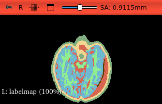
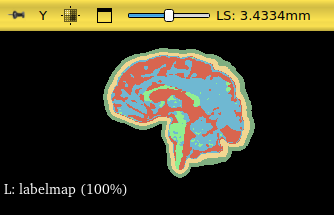
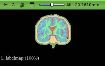
Input
The input is a brain label map where each voxel is assigned a tissue label (e.g., gray matter, white matter, CSF):
Output
The output is a hexahedral mesh of the brain with element attributes corresponding to tissue labels:
Command
mvox --input-attributes labelmap.nrrd --input-tensors cond.nrrd --output-mesh brain.vtu
Data
The data for this example is available at https://zenodo.org/record/7687631.
References
Zwick BF, Bourantas GC, Safdar S, Joldes GR, Hyde DE, Warfield SK, Wittek A, Miller K. Patient-specific solution of the electrocorticography forward problem in deforming brain. NeuroImage. 2022;263:119649. DOI: 10.1016/j.neuroimage.2022.119649
Zwick BF, Safdar S, Bourantas GC, Joldes GR, Hyde DE, Warfield SK, Wittek A, Miller K. Image data and computational grids for computing brain shift and solving the electrocorticography forward problem. Data Brief. 2023;48:109122. DOI: 10.1016/j.dib.2023.109122